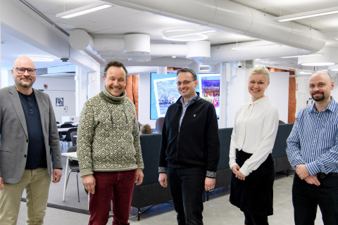
“The brain’s immune cells, microglia, are implicated in virtually all neurological diseases. Particularly Alzheimer’s disease is strongly influenced by the role of microglia and causally linked to microglial dysfunction, as many of the risk genes for AD are expressed by microglia,” said University Professor Christian Madry while visiting the UEF Neuroscience Research Community in Kuopio this week.
During their two-day collaboration visit, Madry, who works at the Institute of Neurophysiology at Charité Universitätsmedizin Berlin, and Professor Christian Haass from LMU Munich and DZNE Munich gave lectures addressing the role of microglia.
As described by Madry, microglia are the brain’s highly versatile immune cells. “Endowed with a wide range of functions to protect the brain, microglia can also adopt detrimental activation states that exacerbate disease progression. Understanding the mechanisms that lead to these different activation states and the transition to harmful phenotypes is a major challenge in current research. Given their influential role in Alzheimer’s disease and other neurodegenerative diseases, novel therapeutic approaches are focusing on the manipulation of microglial function.”
In his lecture, Madry explained how specialised functional profiles of microglia in Alzheimer’s disease are also reflected in distinct electrical membrane properties, which are regulated by the activity of ion channels. He presented his research group’s recent progress in the study of two different types of potassium channels involved in the pathophysiological response of microglia in AD and neuroinflammation. The uptake of amyloid beta and the release of pro-inflammatory cytokines are linked to the function of these channels.
Haass pointed out in his lecture that the amyloid cascade in Alzheimer’s disease is not a hypothesis anymore, but has been clinically verified. What’s more, microglia activities are an integral part of the amyloid cascade, in addition to a parallel and unrelated disease mechanism. Their role in the amyloid cascade is currently being shown in his lab.
“Microglia have long been considered to have detrimental roles in Alzheimer’s disease. However, functional analyses of genes encoding risk factors that are linked to late-onset Alzheimer’s disease, and that are enriched or exclusively expressed in microglia, have revealed unexpected protective functions,” he added.
One of the major risk genes for Alzheimer’s disease is TREM2 where loss-of-function mutations promote the disease. Haass’s research group has shown how protective microglial functions can be boosted by stimulating TREM2 with agonistic antibodies. In his talk he also presented results related to the effects of another AD risk gene, ABI3, on microglia.
Hosted by Professor Mikko Hiltunen who leads the Neuroscience Research Community, as well as Professors Ville Leinonen and Tarja Malm, Haass and Madry also visited the Department of Neurosurgery at Kuopio University Hospital and the electrophysiology core facility at A. I. Virtanen Institute for Molecular Sciences – both involved in a unique research protocol where Alzheimer’s disease is studied using fresh cortical biopsy samples from living NPH patients.
According to Madry, this is all the more important as microglial physiology has mostly been studied on rodent models and cell cultures. “Studies examining microglial function in human brain tissue are extremely sparse, mainly due to limited access to surgically removed brain tissue and difficulties in experimental handling. This is particularly problematic since transcriptomic studies have revealed significant differences in the genetic profile between rodent and human microglia, making research in human tissue indispensable.”
“Clinicians and basic scientists at the University of Eastern Finland have joined forces with a pioneering spirit to enable research on extremely rare neurosurgically resected brain tissue with AD pathology. We hope to collaboratively bundle our expertise and thereby accelerate the translation of findings from animal models to humans,” Madry said.


