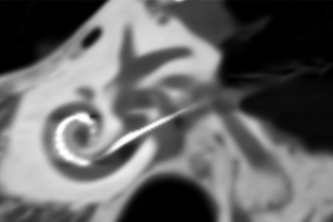
Two newly introduced Cochlear implantation electrodes can provide more atraumatic insertion results, according to the doctoral dissertation of Matti Mustajärvi, Lic Med. The dissertation also introduces new imaging methods for Cochlear implantation follow-up and research.
Cochlear implantation is currently the only routinely used treatment to restore the function of a sense organ. Cochlear implantation was first introduced in 1961 but it was only with the advent of multichannel devices in the early 1990s, that it has gained an established place for the treatment of severe to profound hearing loss.
There are multiple positive and negative factors predicting the hearing outcomes after implantation. One of the most significant negative predictive factors is possible inner ear trauma induced by the surgery. There are mainly two mechanical factors which determine the occurrence of innear ear trauma: electrode design and the insertion technique.
The cone-beam computed-tomography (CB-CT) has recently become a more popular modality in the postoperative evaluation of the results of electrode insertion. The insertion depth, extracochlear electrode contacts, electrode tip fold overs and gross trauma can be easily detected with CB-CT. Even though CB-CT can also quite reliably recognize scala dislocation up to the second turn of the cochlea, a more detailed evaluation of trauma such as elevations or ruptures of the basilar membrane is not possible. Fusion imaging has emerged as a promising modality for achieving a more precise evaluation of electrode positioning and trauma assessment after cochlear implantation.
In the first two studies of this thesis, the insertion results of two newly introduced electrodes were evaluated in freshly frozen temporal bones. The first study was a radiological and histological study that evaluated the Slim Modiolar electrode TM (Cochlear corporation, Sydney, Australia) (SME) which represents a completely new design of a modiolar (precurved) electrode. It was designed to have a more reliable structure and to achieve better hearing preservation than its predecessor, a stylet-type modiolar electrode. In this evaluation study, the researchers detected one scala dislocation in 20 temporal bones inserted with SME. The image fusion with pre- and postoperative CB-CT was performed in all of the 20 TBs. The image fusion proved to be an accurate method in the evaluation of electrode placement inside cochlea.
The second study investigated the insertion characteristics of a new lateral wall electrode, the SlimJ –electrode (Advanced Bionics, Valencia, USA) in 11 freshly frozen temporal bones. In this study, one scala dislocation was found in postoperative fusion imaging. These results are comparable to other temporal bone studies with modern straight electrodes. SlimJ is reasonably predictable with respect to the insertion results, however the final evaluation of insertion properties will require clinical verification.
In the third study, the researchers retrospectively analysed hearing preservation results with SME in 17 clinical patients (18 ears) with low frequency residual hearing. The preliminary results (mean follow-up 582 days) showed a good hearing preservation rate. There were no total hearing losses and seven patients could use electric-acoustic stimulation (EAS). This study revealed significantly more favorable hearing preservation rates than reported for other stylet-type modiolar electrodes.
Fusion imaging was validated with histological samples in the first temporal bone study made with SME. The fusion imaging provided a fast and accurate method for the evaluation of the electrode placement. No significant difference was observed between histologic or fusion imaging measurements.
The fourth study investigated a new fusion imaging technique which may enable better visualization of the basilar membrane. Visualization was conducted in twelwe temporal bones. The perilymph was evacuated from the scalae prior to pre-operative CB-CT. The frozen temporal bone (TB) was initially immersed in Ringer solution to rehydrate both scalae. Insertion was made after rehydration followed by post-operative CB-CT imaging. With the application of this technique, it was easier to detect the individual anatomy of the basilar membrane and a reliable trauma assessment was possible beyond the second turn of the cochlear partition.
The new studied electrode designs provide not only more atraumatic but also more predictable insertion results. The fusion imaging is an accurate method making possible a more detailed electrode placement evaluation as compared to postoperative CB-CT alone. It also represents a fast and cost-effective method for evaluating insertion results in temporal bone studies.
The doctoral dissertation of Matti Iso-Mustajärvi, Licentiate of Medicine, entitled Insertion characteristics of different cochlear implant electrodes: a clinical, radiological and histological study, will be examined at the Faculty of Health Sciences. The Opponent in the public examination will be Professor Jussi Jero of the University of Helsinki, and the Custos will be Professor Heikki Löppönen of the University of Eastern Finland. The public examination will be held online on 15 January 2020 starting at 12 noon.
Dissertation online: Iso-Mustajärvi, Matti. Insertion characteristics of different cochlear implant electrodes: a clinical, radiological and histological study
Image on top: Cone-beam image of inner ear and electrode. Image: Matti Iso-Mustajärvi.

