The doctoral dissertation in the field of Medicine will be examined at the Faculty of Health Sciences at Kuopio campus. The public examination will be streamed online.
What is the topic of your doctoral research? Why is it important to study the topic?
The topic of this dissertation is intraoperative hyperspectral imaging (HSI) of central nervous system tissues. In this dissertation, a microsurgical HSI system and machine learning-based tissue classification methods were developed, and their clinical feasibility was investigated.
Most brain tumour operations are performed microsurgically under high magnification and near vulnerable structures, where injury could lead to severe complications, such as paralysis. Currently, tissue recognition is especially based on anatomical knowledge, visual perception, and indirect feedback from imaging systems. Reliable near real-time identification of healthy and pathological tissue is a central challenge in microsurgery.
Surgical tissue detection techniques are constantly under development both for the identification of normal anatomy and pathological tissues. HSI could significantly improve near real-time tissue classification, but the lack of feasible HSI systems and data limits surgical use. Published microsurgical HSI data is scarce and uniform standards to store and manage medical HSI data do not exist. Producing solutions to these limitations could advance the development of HSI systems, which is expected to advance the effectiveness of surgical treatments significantly.
What are the key findings or observations of your doctoral research?
The dissertation validated the feasibility of HSI in microneurosurgical conditions, and the developed methods serve as the basis for further development of near real-time brain tissue classification systems.
In the preclinical study, machine learning analysis of HSI data differentiated microsurgically critical tissue differences in facial nerves and internal carotid arteries. The method reliably identified microanatomical location- and surgical manipulation-dependent but visually subtle tissue differences.
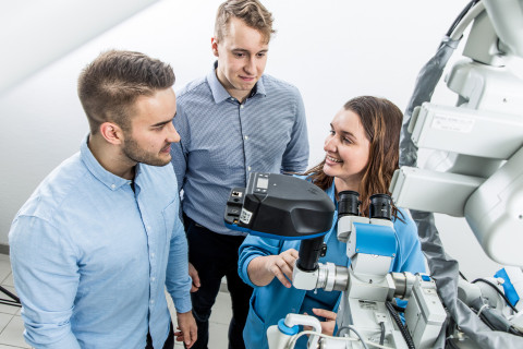
The clinical phase of the dissertation resulted in an operating microscope-mounted HSI system, which was validated for clinical use during brain tumour surgeries.
The system passed the medical device license process and is the only microneurosurgical HSI system in Finland and among the first in Europe. To support the HSI system, we developed a multimodal HSI database that allows clinicians to browse, import, export, and analyse clinical HSI data. In addition to the neurosurgical tissue HSI data, we included relevant clinical information, such as tissue anatomy and relevant MRI scans, which are required to develop intraoperative machine learning HSI applications.
How can the results of your doctoral research be utilised in practice?
The research contributes to the creation of a near real-time tissue classification model, which is applied for the recognition of central nervous tissues and pathologies during surgery. To develop the surgical HSI-solutions further, the system will be applied more broadly, especially for head and neck surgeries.
The HSI systems enable a highly sensitive and individualized tissue classification during surgery. The systems are expected to enhance the accurate recognition of healthy and pathological tissues and the removal of malignant tissue. It would improve surgical performance, help doctors in diagnostics, and thus, diversely promote patient safety. The method requires development before interventional studies, but reported surgical HSI research and our results are promising.
The research data is also utilizable for multidisciplinary innovations and the development of medical imaging systems. The multidisciplinary collaboration of our research group at the Microsurgery Center of Eastern Finland has already produced start-up activity in North Savo: SeeTrue Technologies founded in 2018, and Marginum founded in 2020.
What are the key research methods and materials used in your doctoral research?
In this dissertation, a novel intraoperative tissue classification system based on optical hyperspectral imaging and machine learning was developed. The collected datasets comprised donated human placentas, cadaveric temporal bones, and neurosurgical tissues imaged during brain tumour surgery. The data were analysed using machine learning approaches.
This dissertation is part of research funded by the Finnish Culture Foundation, Finnish Government Research Funding, and Finnish Medical Foundation with docent and neurosurgeon Antti-Pekka Elomaa as the Primary Investigator, as well as the FIRST research project funded by the Finnish Academy with professor Roman Bednarik as the PI. The studies were conducted at the Microsurgery Center of Eastern Finland, the operating rooms of Kuopio University Hospital, and the University of Eastern Finland (UEF) Joensuu campus laboratories. We collaborated with the UEF Color laboratories led by professor Markku Hauta-Kasari, as well as the UEF Department of Physics and Mathematics and HSI-industry representatives, such as the Senop Oy. Our research group has over a decade of multidisciplinary expertise in translative medical device testing following the principles of responsible conduct of research and good clinical practice, and the approval of supervising authorities, such as Valvira and Fimea.
In parallel to the conducted studies, the collected datasets have been used in international courses and master’s theses of around 100 computer science and physics students. The datasets are still being used in UEF computer science courses to solve problems encountered during the development of clinical HSI systems. This collaboration between clinicians, researchers, and students is expected to produce top know-how, ultimately contributing to the development of novel optical imaging solutions for surgeons. These solutions aim to make surgery more efficient, help surgeons avoid complications, and acquire better outcomes.
The doctoral dissertation of Sami Puustinen, Bachelor of Medicine, entitled Intraoperative Hyperspectral Imaging (HSI) in Microneurosurgery: Design for Near Real-Time Optical Tissue Analysis will be examined at the Faculty of Health Sciences. The Opponent in the public examination will be Professor Fady Charbel of University of Illinois Chicago, and the Custos will be Docent Antti-Pekka Elomaa of the University of Eastern Finland.
Doctoral defence
For further information, please contact:
Sami Puustinen, BM, sampuu(a)uef.fi, 0442190767


