ABSTRACT
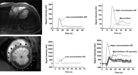
Coronary artery disease (CAD) is the most common cause of death globally, with more than 100 million people suffering from it. Accurate quantification of myocardial blood flow (MBF) is important in diagnostics of CAD and offers important information about the severity and progression of cardiac disease, response to therapy and prognosis. Contrast agent enhanced first-pass MRI (CMR) is a promising method for estimating MBF, because its availability is good, and it does not expose patients to ionizing radiation. This method necessitates monitoring the concentration of contrast agent in blood and myocardium and is based on the assumption that the MRI signal increases linearly with the contrast agent concentration. However, with high contrast agent concentrations this linearity is no more valid. High concentrations may occur in blood pool, where the arterial input function (AIF) for determination of MBF is obtained. Distortion in AIF leads to errors in determined MBF values. Another drawback is related to image artefacts, which are regrettably common in MR imaging. The image artefacts may spoil the MRI signal from myocardium, and therefore impede the accurate estimation of MBF.
In this thesis, a novel method – modified dual bolus method - to correct the high concentration arterial input function (AIF) for determination of MBF was introduced. It was tested with 16 patients in study I and five pigs in study II by comparing the values of Ktrans and MBF with corresponding values determined with the traditional dual bolus method and PET. To solve problems caused by image artefacts, the feasibility of three machine learning methods, support vector machine (SVM), random forest (RF) and linear regression (LR), to estimate the values of MBF from tissue impulse response was investigated in study III. In this analysis, also the artefacted myocardial tissue MRI signals were included. In addition, the capability of SVM and RF methods to recognize the artefacted tissue impulse responses was studied. The performance of these methods were tested with five pigs in rest and stress. The estimated values of MBF were compared with the corresponding values of MBF determined with PET.
Strong correlation in Ktrans values determined with modified dual bolus and traditional dual bolus methods was found in study I, except in cases when heart rate varied significantly during MR imaging. In study II, strong correlation in the values of MBF between modified dual bolus method and PET was seen. The modified dual bolus method was found to give more reliable estimates of MFB than the traditional dual bolus method, especially when the heart rate varied during MRI. In study III, SVM was found to produce the most accurate estimates of MBF. RF method managed also well, but performance of LR method was modest. SVM and RF were able to estimate the values of MBF correctly also for the artefacted areas of myocardium. SVM recognized the artefacted tissue impulse responses most reliably.
To conclude, the modified dual bolus method and SVM and RF machine learning methods improve the reliability in determination of MBF. SVM is also well suited for detecting the artefacted MRI data.
The doctoral dissertation of Lic.Phil. Minna Husso, entitled Quantification of myocardial perfusion using contrast agent enhanced first pass magnetic resonance imaging will be examined at the Faculty of Science and Forestry on the 25th of September. The opponent in the public examination will be Professor Hannu Eskola, Tampere University, and the custos will be Professor Hannu Manninen, University of Eastern Finland.